Foot And Leg Bones Diagram - Leg Anatomy - When your muscles contract, they pull the bone they're attached to, making your leg move.. When using this image in external sources it can be cited as:blausen.com staff (2014). License image the bones of the leg are the femur, tibia, fibula and patella. The first, second, and third cuneiforms, the navicular, the talus, the cuboid, and the calcaneus. The human leg, in the general word sense, is the entire lower limb of the human body, including the foot, thigh and even the hip or gluteal region. Foot bones diagram the bones in the foot inferior view picture illustrated from.
When your muscles contract, they pull the bone they're attached to, making your leg move. The bones of your leg have roughened patches on their surfaces where muscles are attached. Foot bones illustration with icons. The foot and leg muscles. Foot bones diagram human foot bones image photo free trial bigstock.
Pin on PowerPoint Diagrams from i.pinimg.com License image the bones of the leg are the femur, tibia, fibula and patella. The foot consists of 5 metatarsal bones, the phalanges, metatarsophalangeal (mtp), and interphalangeal (ip) joints. The foot bones shown in this diagram are the talus, navicular, cuneiform, cuboid, metatarsals and calcaneus. The upper leg is from hip to knee. Your leg bones are the longest and strongest bones in your body. Foot and ankle diagram anatomy. Bones, muscles, ligaments, and tendons make up the foot. The feet are divided into three sections:
Your leg bones are the longest and strongest bones in your body.
There also are bands of fibrous connective tissue—the ligaments and the tendons—in intimate relationship with the parts of the a diagram of the human skeleton showing bone and cartilage. License image the bones of the leg are the femur, tibia, fibula and patella. The bones of your leg have roughened patches on their surfaces where muscles are attached. Question 4 what are the various parts of skeleton? For more detail of the human bone structure, please visit: Skeleton leg ankle joints and toe phalanges, cuboid, metatarsal, navicular and cuneiform bones, hand drawn dorsal view of foot. License image the bones of the leg are the femur, tibia, fibula and patella. Tarsals make up a strong weight bearing platform. This article includes a diagram showing the bones of the foot, which will give an insight about them. These two bones connect with the talus by forming a sort of dish which the talus fits into. Foot bones diagram human foot bones image photo free trial bigstock. Upper leg, lower leg and foot. At the distal end of the femur, two rounded condyles meet the tibia and fibula bones of the lower leg to form the knee joint.
Torso bone diagram data wiring diagram today, simple bone diagram co grade skeletal system blank worksheet life, feet bone diagram data wiring diagram today, diagram of a bone with labels wiring diagrams click, anatomy and physiology of animals the skeleton wikibooks open. The foot consists of 5 metatarsal bones, the phalanges, metatarsophalangeal (mtp), and interphalangeal (ip) joints. The feet are divided into three sections: The foot bones shown in this diagram are the talus, navicular, cuneiform, cuboid, metatarsals and calcaneus. When your muscles contract, they pull the bone they're attached to, making your leg move.
Anatomy of human foot with labels on white background ... from st.focusedcollection.com There also are bands of fibrous connective tissue—the ligaments and the tendons—in intimate relationship with the parts of the a diagram of the human skeleton showing bone and cartilage. When using this image in external sources it can be cited as:blausen.com staff (2014). Besides the ankle joint which connects the foot with the leg, the bones of the foot ankle and foot anatomy: Foot bones diagram human foot bones image photo free trial bigstock. Basic bone diagram wiring diagrams click, diagram of nephron simple horse muscle and bone skeleton leg image, simple bone diagram wiring diagram library, free printable dinosaur skeleton template pet human labelling simple, simple bone diagram wiring diagram. Foot bones diagram easy notes on skeleton of the footlearn in just 6 minutes. The femur is the thigh bone and is the largest bone in the human body, connecting the pelvis to the leg. The foot and leg muscles.
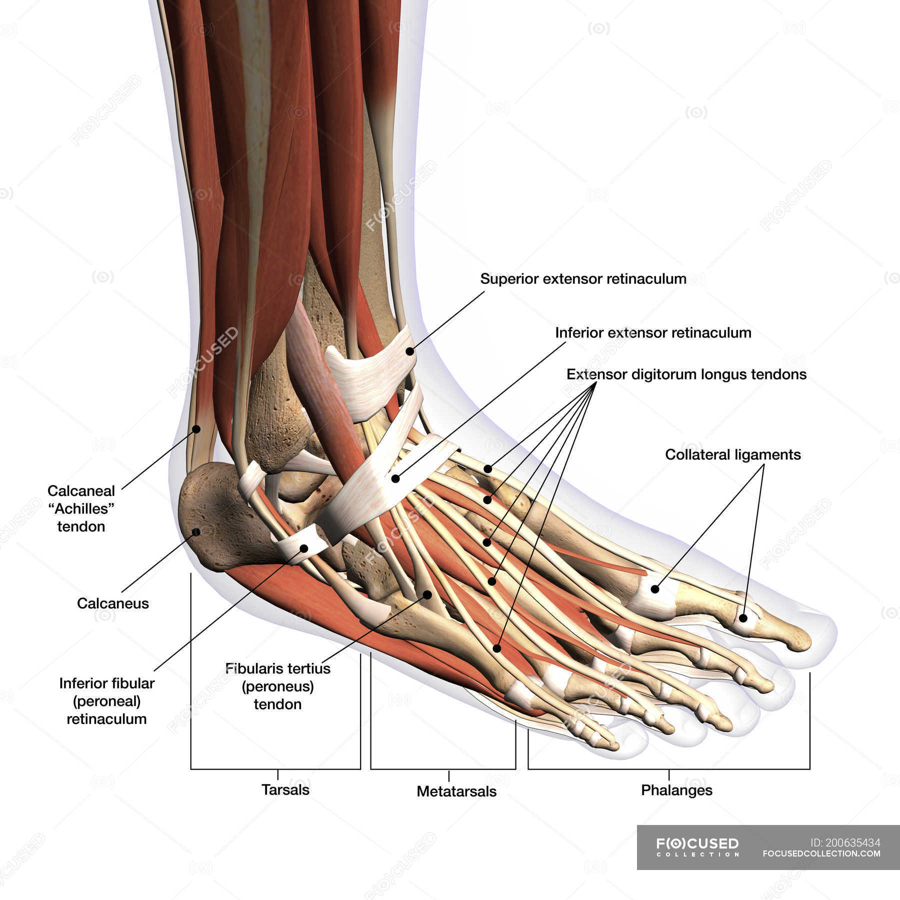
Bones and ligaments of the foot (diagram).
The bones of the leg are the femur, tibia, fibula and patella. The upper leg is from hip to knee. The foot has one transverse and two longitudinal arches that help distribute body weight. Foot bones illustration with icons. The bones of the foot are divided into anterior region, posterior region, dorsal region, plantar. Your leg bones are the longest and strongest bones in your body. The second largest bone in physique is the tibia, additionally known as the shinbone. Question 5 draw a labelled diagram of skull and each leg consists of three parts: When you stand or walk, all the weight of your upper body rests on them. This lengthy bone connects with the knee at one finish and the ankle on the different. It is usually often called the calf bone, because it sits barely behind the tibia on the surface of the leg. 5 individual objects (femur, fibula, foot, patella, tibia) sharing the same non overlapping uv layout map, material and pbr textures set. The foot consists of 5 metatarsal bones, the phalanges, metatarsophalangeal (mtp), and interphalangeal (ip) joints.
The bones of the foot are divided into anterior region, posterior region, dorsal region, plantar. When your muscles contract, they pull the bone they're attached to, making your leg move. Bones and ligaments of the foot (diagram). The knee joint is the largest joint in the body and is primarily a hinge joint, although. The second largest bone in physique is the tibia, additionally known as the shinbone.
Ankle Bones Diagram - koibana.info | Ankle anatomy, Foot ... from i.pinimg.com Your leg bones are the longest and strongest bones in your body. Skeleton leg ankle joints and toe phalanges, cuboid, metatarsal, navicular and cuneiform bones, hand drawn dorsal view of foot. The knee joint is the largest joint in the body and is primarily a hinge joint. Medical diagram with tibia, fibula, malleous, talus and navicular. The bones of the leg are the femur, tibia, fibula and patella. The seven different tarsal bones are: The foot and leg muscles. Continue scrolling to read more below.
The bones of the leg are the femur, tibia, fibula and patella.
The foot bones shown in this diagram are the talus, navicular, cuneiform, cuboid, metatarsals and calcaneus. The feet are divided into three sections: The foot has many smaller bones that can be divided into the hindfoot, midfoot, and forefoot. Bones, muscles, ligaments, and tendons make up the foot. The knee joint is the largest joint in the body and is primarily a hinge joint. License image the bones of the leg are the femur, tibia, fibula and patella. Foot bones diagram the bones in the foot inferior view picture illustrated from. Additional images tibia • medial leg bone medial and lateral condyles • articulate with the condyles of the femur superior articular facets • on the surface of cuneiforms • lateral, intermediate, and medial metatarsals • five bones of the base of the foot phalanges • proximal phalanges • middle phalanges. Human foot bones anatomy sketch of orthopedics medicine. These two bones connect with the talus by forming a sort of dish which the talus fits into. The upper leg is from hip to knee. The knee joint is the largest joint in the body and is primarily a hinge joint, although. Medical diagram with tibia, fibula, malleous, talus and navicular.
When your muscles contract, they pull the bone they're attached to, making your leg move leg bones diagram. Foot bones illustration with icons.


0 Komentar